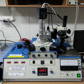
Precision Ion Polishing System (Gatan PIPS I – Model 691)
The PIPSTM ion mill is a user-friendly, tabletop Precision Ion Polisher System designed to produce high quality, TEM specimens with large electron transparent areas. The PIPSTM incorporates patented Whisperlok® stage, 2 unique penning ion guns with +10˚ to -10˚ milling angles, variable energy milling (down to 100 eV), liquid nitrogen specimen cooling and an oil-free vacuum system for ultra-clean specimen processing. (quoted from Gatan, inc.)Argon Ion Source
-Milling angle: +10˚ to -10˚
- Ion beam energy: 100 eV to 6.0 keV
- Ion current density: 10 mA/cm2 peak
- Beam diameter: 350 to 800 ㎛ FWHM at 5 keV
- Beam modulation: Single or Double sector LN2 Cold Stage Installed Real-time Video Monitoring System Installed (Acquired in 2008)
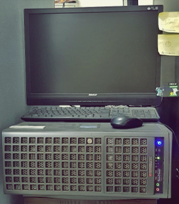
GPU Cluster
Transmission electron microscopy has been undergoing a transition; due to advances in technologies such as 4D STEM, 3D tomography, and STEM EELS, it is now possible to obtain an abundant amount of data at once. Therefore, in recent transmission electron microscopy, ways to process such a huge amount of data is a crucial issue. GPU clusters are utilized to perform various data processing. For instance, noise reduction by principal component analysis, 3D tomography reconstruction, and fluctuation electron microscopy are being studied.-20 cores 2.2GHz CPU x2
-128GB 2666 MHz RAM
-112 teraFLOPS 32GB GPU x 2
-1TB SSD storage
-12TB HDD RAID 1 storage
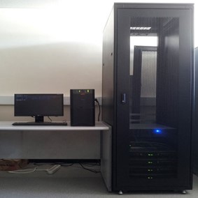
Parallel Computer Cluster
Parallel computer clusters are utilized to calculate electronic structures based on first-principles density functional theory. Nowadays, density functional theory is used extensively to complement microscopic data and has successfully bridged atomic structure to macroscopic properties in many cases. Combining HR-STEM image with density functional theory, we can estimate physical and chemical properties stemmed from its own atomic structure of materials. Also, Combining the experimental EELS with the theoretical DFT calculations, we can perform the simulation of experimental data and give good explanation for the spectrum. Thus, active utilization of density functional theory with STEM/EELS experiments can be great helpful to understand the novel properties in materials.4-node hexa core processor cluster
Density functional theory simulation package
-VASP
-WIEN2k
Dynamic electron scattering simulation
-Zmult
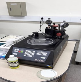
MultiPrep Polishing System (Allied 15-2000 MultiPrep)
The MultiPrep system enables precise semi-automatic sample preparation of a wide range of materials for microscopic evaluation. Capabilities include parallel polishing, angle polishing, site-specific polishing or any combination thereof. Dual micrometers allow precise sample tilt adjustments relative to the abrasive plane. A rigid Z-indexing spindle maintains the pre-defined geometric orientation throughout the grinding/polishing process. Digital indicators enable quantifiable material removal, which can be monitored real-time, or pre-set for unattended operation. Variable speed rotation and oscillation maximize use of the entire abrasive/polishing disc and minimize artifacts.8" polishing system
- 8“ diamond lapping film
- 8“ red final C polishing cloth
Mitutoyo absolute digimatic indicator
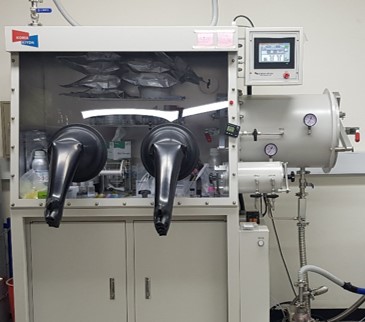
Glove box(KK-011AS)
The glove box is used for battery research. Materials used in battery research should be implemented in a controlled atmosphere.Purifity Level: Less than 1 ppm of oxygen and moisture concentration level
Catalyst : Oxygen Catalyst and Molecular Sieve
PLC: Automatic PLC control
Control Panel: 7”Full Color Touch Screen
Purifier Regeneration/: Auto Regeneration programmed on PLC
Blower Speed Control: Max. 63 CFM (1.78m3/min)
Vacuum Pump: 200L/min
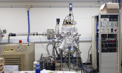
Molecular Beam Epitaxy system
An ultrahigh vacuum molecular beam epitaxy (UHV-MBE) system is designed for topological insulator and superconductor van der Waals materials growth.Source : Gallium, Selenium
Shared Facilities
Following TEM instruments are owned by and located in Research Institute of Advanced Materials (RIAM)
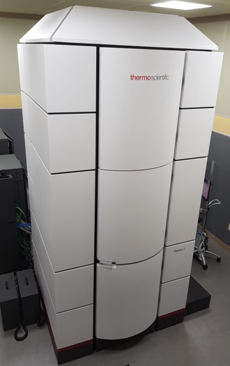
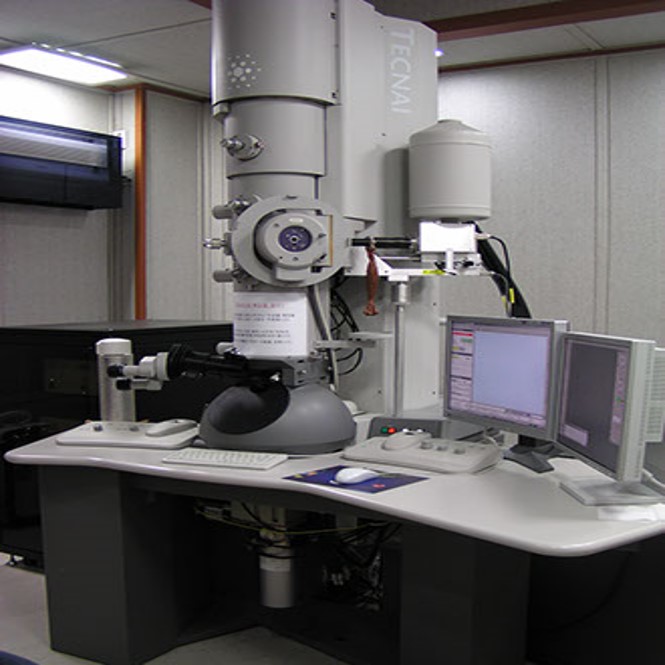
Cs corrected monochromated TEM/STEM (Themis Z)
Accelaration voltage : 60 -300kV (60, 80, 200, 300)Resolution
- Image corrector : Information Limit 70pm, STEM resolution 136pm
- Probe corrector : Information Limit 100pm, STEM resolution 60pm
- X-FEG/ monochromator+image corrector : Information Limit 60pm, STEM resolution 60pm
EDS energy resolution at Mn ≤ 136eV (all detectors) : 133.4eV
Monochromator energy resolution
: 300kV : 0.17eV, 200kV : 0.14eV, 80kV : 0.17eV, 60kV : 0.16eV
HR-STEM resolution (HAADF with S-CORR≤0.06 nm) : 0.05nm
Analytic accessaries :
Atomic-Resolution (S)TEM imaging Analysis
Chemical analysis using by Monochromated EELS & EDS
4D STEM using by Pixel Array 4D STEM Detector
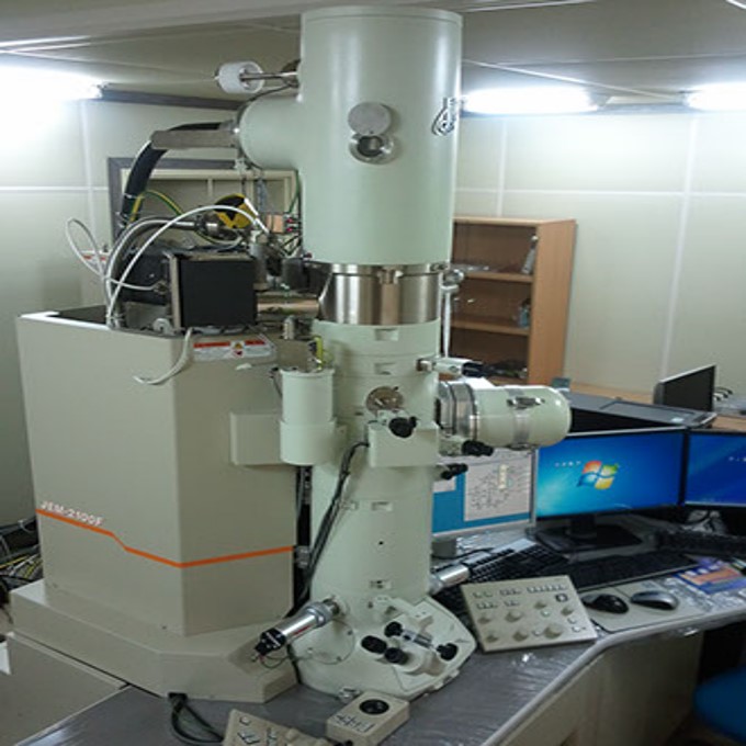
JEM-2100F
Accelaration voltage : 200kVGun : Field Emission Gun
Point resolution : 2.3Å
Line resolution : 1.0Å
STEM BF lattice resolution: 2 Å
Magnification: x50 ~ x1500,000
Analytic accessaries
EDS (Energy Dispersive Spectroscopy)
STEM (Scanning Transmission Electron Microscopy)
HAADF (High Angle Annular dark Field)
DIGISTAR (Orientation-Phase imaging with Precession Diffraction)
Tecnai F20
Accelaration voltage : 200kVGun : Field Emission Gun
Point resolution : 2.5Å
Line resolution : 1.02Å
Magnification: x22 ~ x930,000
Analytic accessaries
EDS (Energy Dispersive Spectroscopy)
EELS (Electron Energy Loss Spectroscopy
HAADF (High Angle Annular dark Field)
DIGISTAR (Orientation-Phase imaging with Precession Diffraction)
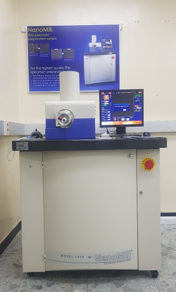
NanoMill(Model 1040)
Fischione’s Model 1040 NanoMill® TEM specimen preparation system is a tool for creating the high-quality thin specimens needed for advanced transmission electron microscopy imaging and analysis. It is ideal for both post-FIB (focused ion beam) processing and the enhancement of conventionally prepared specimens. The NanoMill system features gaseous ion source technology that results in ion energies as low as 50 eV and a beam size as small as 1 µm. It allows specimens to be prepared without amorphization, implantation, or redeposition. The ion beam can be targeted to a specific area of interest. A secondary electron detector is used to image the ion-induced secondary electrons that are generated from the targeted area of the specimen. (quoted from Fischione, inc.)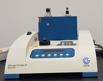
Dimple Grinder II
Dimple grinder enables fast and reliable mechanical pre-thinning to near electron transparency to greatly reduce your ion milling times and uneven thinning. (quoted from Gatan, inc.)